Anúncios
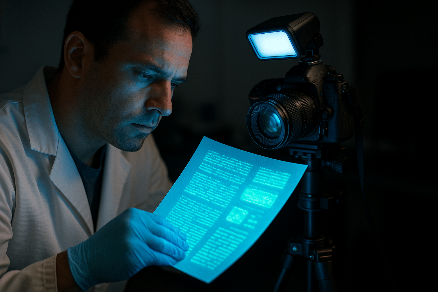
Fluorescence imaging has revolutionized scientific research, enabling visualization of cellular processes with unprecedented clarity and precision in modern laboratories worldwide.
🔬 The Foundation of Fluorescence Documentation
Fluorescence microscopy stands as one of the most powerful tools in biological research, allowing scientists to observe molecular interactions, cellular structures, and dynamic processes in real-time. The ability to document these phenomena with accuracy requires not just sophisticated equipment, but a comprehensive understanding of imaging principles and best practices.
Anúncios
Modern research demands more than simple observation. Scientists need to capture, quantify, and analyze fluorescent signals with reproducibility and precision. This requirement has driven innovations in camera technology, software solutions, and imaging protocols that collectively transform raw fluorescent signals into meaningful scientific data.
The journey from sample preparation to publication-ready images involves multiple critical steps. Each phase presents unique challenges that can impact the quality and reliability of your research outcomes. Understanding these challenges and implementing appropriate solutions separates mediocre documentation from exceptional scientific imaging.
Understanding the Science Behind Fluorescent Signals
Fluorescence occurs when molecules absorb light at specific wavelengths and emit it at longer wavelengths. This phenomenon, discovered over a century ago, has become the backbone of cellular and molecular biology. The key to successful fluorescence documentation lies in maximizing signal detection while minimizing noise and photobleaching.
Anúncios
Different fluorophores exhibit distinct excitation and emission spectra, requiring careful selection based on experimental requirements. Common fluorescent proteins like GFP, RFP, and their variants offer researchers a palette of colors for multi-label experiments. Synthetic dyes such as Alexa Fluor, FITC, and rhodamine provide alternatives with superior brightness and photostability.
The intensity of fluorescent signals depends on numerous factors including fluorophore concentration, quantum efficiency, excitation light intensity, and the optical properties of your microscope system. Environmental conditions like pH, temperature, and oxygen levels also influence fluorescence behavior, making controlled experimental conditions essential.
Critical Parameters for Image Acquisition
Achieving precision in fluorescence imaging requires careful attention to several technical parameters. Exposure time determines how long the camera sensor collects photons from your sample. Too short, and you’ll miss weak signals; too long, and you risk photobleaching and saturated pixels that destroy quantitative accuracy.
Gain settings amplify the signal from your camera sensor but also amplify background noise. Finding the optimal balance ensures you capture genuine fluorescent signals without introducing artifacts. Modern scientific cameras offer various gain modes optimized for different signal intensities and experimental conditions.
Binning combines adjacent pixels to increase sensitivity and reduce file size, but sacrifices spatial resolution. This trade-off becomes critical when imaging dim samples or conducting time-lapse experiments where speed matters. Understanding when to implement binning requires balancing your specific experimental needs against image quality requirements.
🎯 Equipment Selection for Optimal Results
The foundation of accurate fluorescence documentation begins with appropriate hardware selection. Scientific-grade cameras designed specifically for fluorescence imaging offer superior sensitivity, dynamic range, and quantum efficiency compared to standard photography equipment.
CCD and sCMOS cameras represent the gold standard for fluorescence imaging. CCD sensors traditionally offered excellent sensitivity and low noise, particularly in cooled configurations. sCMOS technology has evolved to provide comparable sensitivity with faster readout speeds, making them ideal for dynamic imaging applications.
Your microscope system’s optical components significantly impact image quality. High numerical aperture objectives collect more light, improving signal strength and resolution. Quality filters with sharp spectral edges prevent excitation light bleed-through while maximizing emission signal transmission. Light sources must provide stable, uniform illumination across the field of view.
Illumination Strategies That Make the Difference
LED illumination has largely replaced traditional mercury and xenon arc lamps in modern fluorescence microscopy. LEDs offer instant on-off switching, eliminating warm-up times and reducing photobleaching between acquisitions. Their narrow spectral output increases contrast by reducing out-of-band excitation.
Laser-based systems provide the highest light intensity and spectral purity, essential for confocal and super-resolution techniques. The coherent nature of laser light enables precise focusing and scanning, though it can introduce speckle patterns that require mitigation strategies.
Illumination intensity directly affects both signal strength and phototoxicity. Too much excitation light damages living cells and accelerates photobleaching. Too little compromises image quality and extends exposure times. Implementing intelligent control systems that deliver just enough light for each application optimizes both image quality and sample viability.
Mastering Image Acquisition Protocols
Standardized imaging protocols ensure reproducibility across experiments and laboratories. Begin by establishing baseline settings with test samples, documenting every parameter for future reference. This systematic approach eliminates guesswork and enables meaningful comparisons between experimental conditions.
Background correction removes inherent noise from your imaging system. Capture dark frames with identical settings but no illumination to measure camera noise. Flat-field correction compensates for uneven illumination across the field of view, ensuring accurate intensity measurements throughout your images.
Multi-channel imaging requires sequential or simultaneous acquisition of different fluorophores. Sequential imaging prevents spectral bleed-through but may miss rapid dynamic events. Simultaneous acquisition captures synchronized information but requires careful spectral separation to avoid cross-talk between channels.
Time-Lapse Considerations for Dynamic Processes
Time-lapse fluorescence imaging reveals dynamic cellular processes but demands careful planning. Frame rate must balance temporal resolution against photobleaching and phototoxicity. Too frequent imaging damages cells; too infrequent imaging misses critical events.
Environmental control becomes paramount during extended time-lapse experiments. Temperature, humidity, and CO2 levels must remain stable to maintain cell viability. Stage drift over hours or days can shift your region of interest out of the field of view, requiring autofocus systems and stage stabilization.
Data management challenges escalate quickly during time-lapse experiments. Multi-dimensional datasets spanning time, multiple channels, and z-stacks can generate hundreds of gigabytes. Implement efficient file formats and storage strategies before beginning acquisition to prevent bottlenecks during critical experiments.
📊 Quantitative Analysis and Data Integrity
Transforming fluorescence images into quantitative data requires rigorous analysis methods. Intensity measurements must account for background fluorescence, photobleaching over time, and variations in sample thickness. Proper calibration against known standards establishes absolute measurements rather than arbitrary units.
Colocalization analysis determines whether different fluorophores occupy the same cellular locations, revealing protein interactions and compartmentalization. Pearson’s correlation coefficient and Manders’ coefficients provide statistical measures of colocalization, but require appropriate threshold settings and background subtraction.
Particle tracking algorithms follow individual molecules or organelles through time-lapse sequences, quantifying movement patterns, velocities, and diffusion coefficients. These measurements reveal fundamental biological processes like protein trafficking, vesicle transport, and cell migration.
Software Solutions for Professional Analysis
ImageJ and Fiji represent powerful, open-source platforms for fluorescence image analysis. Their extensive plugin ecosystems provide specialized tools for virtually every analysis need. From basic intensity measurements to advanced machine learning-based segmentation, these platforms serve researchers worldwide.
Commercial software packages offer refined user interfaces, automated workflows, and technical support. Platforms like MetaMorph, NIS-Elements, and Imaris excel at specific tasks such as 3D reconstruction, object tracking, and multi-dimensional dataset management. The choice between open-source and commercial solutions depends on your specific requirements and budget constraints.
Machine learning algorithms increasingly enhance fluorescence image analysis. Deep learning models trained on annotated datasets can segment complex structures, classify cellular phenotypes, and detect subtle patterns invisible to human observers. These AI-powered tools accelerate analysis while reducing subjective bias.
🛡️ Preventing Common Pitfalls and Artifacts
Photobleaching remains one of the most significant challenges in fluorescence imaging. This irreversible destruction of fluorophores limits observation time and compromises quantitative accuracy. Anti-fade mounting media, reduced illumination intensity, and optimized acquisition protocols minimize photobleaching effects.
Autofluorescence from biological samples creates background signals that obscure genuine fluorescent labels. Different tissues and cellular components exhibit varying levels of autofluorescence. Spectral unmixing techniques can separate autofluorescence from specific fluorophore signals, though prevention through careful fluorophore selection remains preferable.
Out-of-focus light degrades image contrast in widefield fluorescence microscopy. Deconvolution algorithms mathematically remove blur based on the microscope’s point spread function, restoring clarity to three-dimensional datasets. Confocal microscopy physically eliminates out-of-focus light through pinhole apertures, though at the cost of reduced light efficiency.
Calibration and Quality Control Essentials
Regular calibration maintains imaging system performance over time. Fluorescent beads with known spectral properties and intensities serve as standards for verifying excitation alignment, emission collection efficiency, and system linearity. Document calibration results to track performance trends and identify degrading components.
Resolution test targets reveal your system’s true spatial resolution, often lower than theoretical limits due to optical aberrations or misalignment. Regularly measuring resolution ensures your images contain genuine detail rather than artifacts masquerading as structure.
Intensity calibration converts arbitrary camera units into absolute physical measurements. Fluorescent standards with certified brightness enable comparisons across different microscopes, experiments, and laboratories. This standardization proves essential for quantitative biology and reproducible science.
Advanced Techniques for Specialized Applications
Super-resolution microscopy breaks the diffraction limit, revealing structures smaller than 200 nanometers. STED, PALM, STORM, and structured illumination microscopy each offer unique advantages for different applications. These techniques require specialized equipment and sample preparation but provide unprecedented detail of cellular architecture.
Fluorescence lifetime imaging (FLIM) measures how long fluorophores remain in the excited state before emitting photons. This parameter depends on molecular environment rather than concentration, providing information about pH, ion concentrations, and protein-protein interactions invisible to intensity-based measurements.
Light sheet microscopy illuminates samples from the side while imaging from above, dramatically reducing photobleaching and phototoxicity. This approach enables long-term imaging of living embryos, tissue cultures, and even whole organisms, opening new windows into developmental biology and disease progression.
💡 Optimizing Workflow Efficiency
Efficient workflows save valuable research time while improving data quality. Automated stage control and position memory enable high-throughput screening of multiple samples or experimental conditions. Pre-programmed acquisition sequences ensure consistent imaging parameters across all samples.
Metadata management becomes crucial as datasets grow complex. Recording all acquisition parameters, sample details, and experimental conditions within image files ensures traceability and enables future reanalysis. Standardized naming conventions and folder structures prevent confusion and data loss.
Image processing pipelines automate repetitive analysis tasks, reducing human error and increasing throughput. Batch processing applies identical operations to entire datasets, ensuring consistency. Macro recording features in analysis software capture manual operations for automated replay.
Documentation and Reproducibility Standards
Publishing fluorescence images requires adherence to community standards. The Microscopy Image Data (MIDA) format and similar initiatives promote complete metadata reporting. Journal requirements increasingly demand raw data deposition and detailed methodology descriptions enabling independent reproduction.
Figure preparation must balance aesthetic appeal with scientific integrity. Linear adjustments to brightness and contrast are acceptable, but any non-linear manipulations require explicit disclosure. Consistent lookup tables across experimental conditions prevent misleading visual impressions.
Statistical considerations apply to microscopy as to other experimental methods. Sample sizes must provide adequate statistical power. Blind analysis prevents unconscious bias during quantification. Appropriate statistical tests validate conclusions drawn from imaging data.
Future Directions in Fluorescence Imaging Technology
Emerging technologies continue expanding fluorescence imaging capabilities. Adaptive optics correct aberrations introduced by thick biological samples, maintaining resolution deep within tissues. These systems originally developed for astronomy now enhance biological imaging through wavefront correction.
Novel fluorophores with improved properties appear regularly. Brighter probes enable detection of scarce molecules. Red-shifted fluorophores penetrate deeper into tissues with reduced scattering and autofluorescence. Photoactivatable variants enable temporal control of labeling for sophisticated tracking experiments.
Integration with complementary techniques yields comprehensive information about biological systems. Correlative light and electron microscopy combines fluorescence specificity with ultrastructural detail. Combined fluorescence and atomic force microscopy reveals mechanical properties alongside molecular distributions.

🎓 Building Expertise Through Practice and Community
Mastering fluorescence imaging requires continuous learning and practice. Online courses, workshops, and conferences provide training opportunities at all levels. Manufacturers offer application notes and webinars demonstrating best practices with their equipment.
Core facility staff possess extensive expertise developed through diverse projects and troubleshooting experiences. Collaboration with facility personnel accelerates learning and helps avoid costly mistakes. Many facilities offer training programs tailored to specific techniques or applications.
The scientific imaging community actively shares knowledge through online forums, social media groups, and publications. Engaging with these communities provides solutions to specific problems and keeps researchers updated on emerging techniques and technologies.
Precision fluorescence imaging represents both art and science, requiring technical knowledge, attention to detail, and creative problem-solving. As biological questions grow increasingly complex, imaging capabilities must evolve to match. By mastering fundamental principles, implementing rigorous protocols, and embracing new technologies, researchers transform fluorescence from a simple visualization tool into a quantitative platform that illuminates the molecular mechanisms of life. The investment in proper technique and equipment pays dividends through reliable, reproducible data that advances scientific understanding and earns peer recognition in this competitive field.

